
Endoscopy results usually describe what the camera saw, what tissue samples were taken, and whether follow-up testing is needed. Common findings include a normal exam, irritation, reflux-related changes, gastritis, ulcers, hiatal hernia, polyps, or biopsy results still pending. Ask the clinician what was seen, biopsied, and recommended next.
How did we evaluate endoscopy results?
We evaluated endoscopy results by separating the procedure note, visual findings, pathology report, medication plan, and follow-up instructions. Gastroenterology society guidance, government patient resources, and hospital-based explanations received more weight than forum anecdotes because endoscopy words can sound alarming without context. We prioritized terms people commonly see after upper endoscopy, including normal mucosa, esophagitis, gastritis, hiatal hernia, Barrett’s esophagus, ulcer, polyp, biopsy, and H. pylori. We excluded product recommendations because a cold-stage results article should help readers understand the report and prepare questions, not push a supplement or brand. The limitation is important: an online article cannot interpret an individual pathology result, photo, or medication history. The safest reading combines the written report, pathology addendum, symptom pattern, age, risk factors, medication exposure, and the clinician’s actual follow-up plan. Unclear phrases deserve clinician translation before a reader changes habits.
What does an endoscopy report usually include?
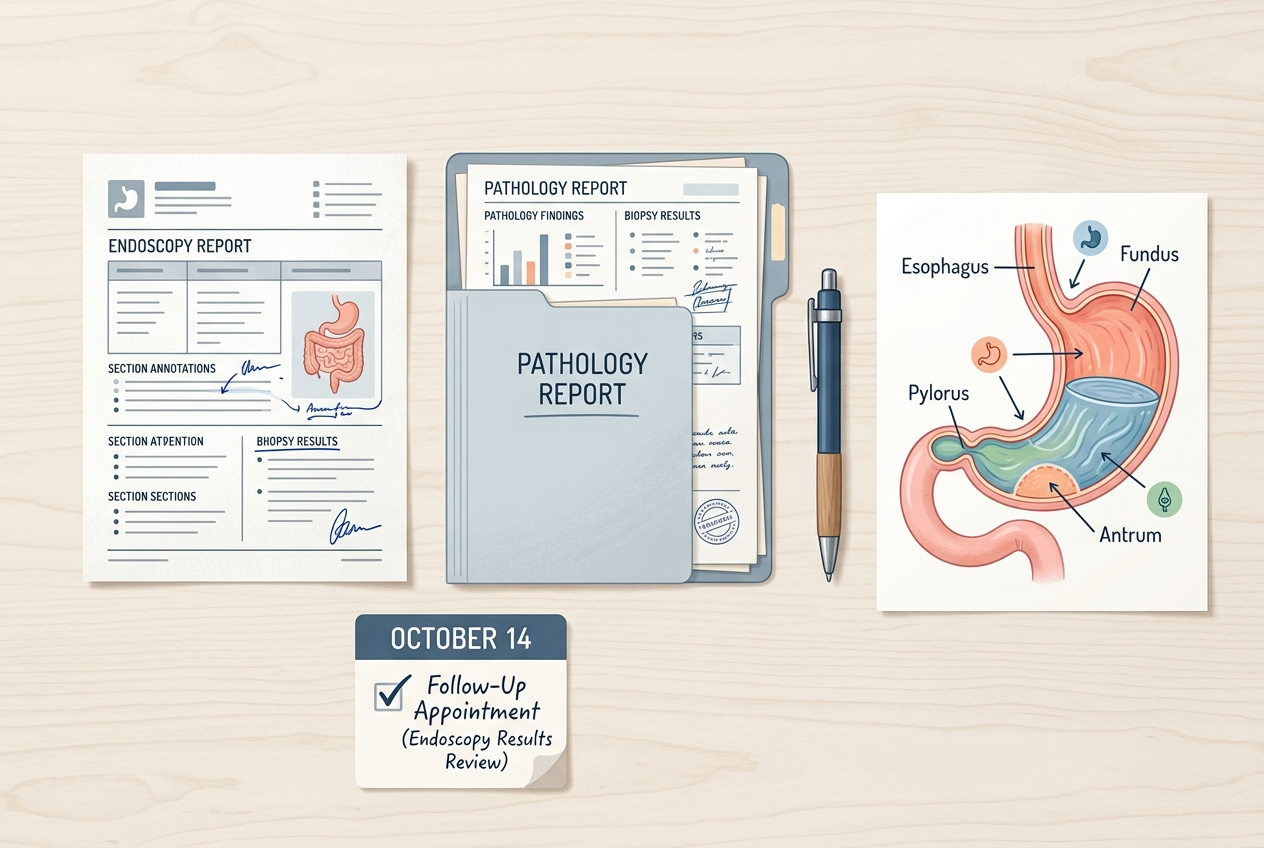
An upper endoscopy report usually includes the indication, sedation details, areas examined, visual findings, biopsy locations, immediate impression, and follow-up plan. The NIDDK upper GI endoscopy guide explains that the test lets a doctor view the esophagus, stomach, and duodenum and may include biopsy or treatment during the same procedure. The indication tells why the test happened, such as trouble swallowing, reflux symptoms, anemia, bleeding signs, nausea, abdominal pain, or follow-up of a prior finding. The findings section describes what the endoscopist saw with the camera. The pathology report describes what a lab saw under a microscope if tissue samples were taken. Those two parts can differ. A camera can show redness, but pathology can identify inflammation type, infection clues, metaplasia, or normal tissue. People should wait for both parts before treating the first result message as the full answer.
What does a normal endoscopy result mean?
A normal upper endoscopy means the endoscopist did not see obvious structural damage in the inspected esophagus, stomach, or duodenum. It does not mean symptoms are imaginary, and it does not rule out every digestive explanation. Functional dyspepsia, reflux without visible erosions, motility problems, food intolerance patterns, medication irritation, constipation pressure, gallbladder disease, and gut-brain interaction can still create real symptoms after a normal exam. The American College of Gastroenterology patient page on functional dyspepsia describes persistent upper-abdominal symptoms that can occur without an ulcer or visible explanation on endoscopy. A normal result can still be useful because it reduces concern for some structural problems and guides next-step testing. The key question is what the clinician thinks fits the whole case now. Ask whether biopsy results are pending, whether H. pylori was checked, and what symptom pattern should trigger follow-up.
What do gastritis, esophagitis, and irritation mean?
Gastritis means the stomach lining shows inflammation or irritation, while esophagitis means the esophagus shows inflammation or injury. These terms describe tissue appearance; they do not automatically explain the cause. Possible contributors include reflux exposure, H. pylori infection, NSAID use, bile irritation, alcohol exposure, autoimmune patterns, or other medical contexts. The Mayo Clinic gastritis overview notes that gastritis can be sudden or gradual and can have different causes. Endoscopy language such as “mild,” “erosive,” “non-erosive,” “erythema,” or “friability” helps classify appearance, but biopsy and history often decide the meaning. Ask whether the finding was mild, moderate, or severe; whether ulcers were present; whether biopsies were taken; and whether medications, infection testing, or follow-up endoscopy were recommended. A short report phrase should not be read as the whole diagnosis.
What does a biopsy after endoscopy mean?
A biopsy after endoscopy means the clinician removed tiny tissue samples for lab review. Biopsy does not automatically mean cancer, and it is common during upper endoscopy when clinicians need to check inflammation, H. pylori, celiac-related changes, Barrett’s esophagus, eosinophilic esophagitis, ulcers, polyps, or tissue that looked different from expected. The pathology report may arrive days after the procedure note, so the first portal message can be incomplete. A useful question is, “Were biopsies routine, targeted, or both?” Routine biopsies sample normal-looking tissue for microscopic clues. Targeted biopsies sample a visible area such as an ulcer edge, polyp, patch, ring, or abnormal lining. The final meaning depends on the microscopic diagnosis, margin comments if relevant, and clinician interpretation. People should save the pathology report, not only the endoscopy photo page, because future clinicians often need the exact wording later.
What does a hiatal hernia on endoscopy mean?
A hiatal hernia means part of the stomach sits higher through the diaphragm opening than usual. Small sliding hiatal hernias are common findings, and their importance depends on size, reflux pattern, symptoms, esophagitis, swallowing issues, anemia clues, and the clinician’s exam. Cleveland Clinic explains that hiatal hernia can be associated with reflux symptoms, but the finding can also be mild or incidental in some people (Cleveland Clinic). The report may describe the hernia in centimeters or by landmarks such as the gastroesophageal junction and diaphragmatic pinch. Ask whether the hernia was small, sliding, paraesophageal, or clinically significant. Also ask whether it changes the reflux plan, eating pattern advice, medication timing, or need for additional testing. The word “hernia” sounds dramatic, but the management question depends on anatomy plus symptoms, not the word alone.
What questions should you ask after getting results?
The best follow-up questions turn report language into next steps. Ask: “What was seen visually, what was biopsied, and what is still pending?” Then ask whether the finding explains the symptoms, whether H. pylori or celiac testing was done, whether medication timing should change, and what warning symptoms should trigger urgent care. If the report mentions esophagitis, ask for the grade. If it mentions Barrett’s esophagus, ask whether intestinal metaplasia was confirmed by pathology and what surveillance interval applies. If it mentions ulcers, ask about NSAIDs, H. pylori, bleeding risk, and follow-up. If it says normal, ask what non-structural explanations remain. Bring a medication list, supplement list, symptom timeline, stool changes, weight changes, and family history to the visit. The goal is not to memorize medical vocabulary; the goal is to know the next decision.
What questions do people ask about endoscopy results?
Can an endoscopy be normal even if symptoms are severe?
Yes. Severe symptoms can occur with a normal-looking endoscopy when the cause is functional dyspepsia, reflux without erosions, visceral sensitivity, motility issues, constipation pressure, or a non-upper-GI source. A normal result should lead to a next-step plan, not dismissal.
Does a biopsy mean the doctor found something bad?
No. Biopsies are common and often routine during endoscopy. The lab may check for inflammation, H. pylori, celiac-type changes, eosinophils, Barrett’s-related changes, or other microscopic findings that the camera cannot confirm alone.
How long do pathology results take?
Timing varies by lab and health system, but pathology commonly takes several business days. Ask the endoscopy office when to expect the final report and whether the clinician will call or post results in the patient portal.
What does mild gastritis mean?
Mild gastritis means the stomach lining looked mildly inflamed or irritated, but the cause and importance depend on biopsy, medications, alcohol exposure, H. pylori testing, and symptoms. Ask whether the finding changes treatment or simply explains part of the pattern.
Can endoscopy diagnose reflux?
Endoscopy can identify reflux complications such as erosive esophagitis, strictures, or Barrett’s changes, but reflux can exist without visible erosions. Some people need symptom review, medication response, pH testing, or other evaluation depending on the case.
What should I do if I do not understand the report?
Write down the exact phrases that confuse you and ask the clinician to translate each one into significance, cause, and next step. Portal summaries can be brief; a follow-up conversation often explains what the wording means for your situation.
What is the bottom line?
Endoscopy results are most useful when read in layers: visual findings first, pathology second, clinician interpretation third, and follow-up plan last. A normal report can still leave real symptoms to solve. An abnormal phrase can be mild, routine, urgent, or simply incomplete until pathology returns. The practical next step is to ask what was seen, what was sampled, what is pending, and what should happen next. Save both the endoscopy note and the pathology report because future clinicians may need exact wording. If symptoms worsen, swallowing becomes difficult, bleeding signs appear, or weight loss is unexplained, contact the care team instead of waiting for the next routine appointment. The report is a map for follow-up, not a final self-diagnosis. Good interpretation turns medical vocabulary into actions: monitor, test, adjust, return, or escalate appropriately afterward.
Leave a Reply