CT, ultrasound, and endoscopy answer different digestive questions. CT shows cross-sectional abdominal structures, ultrasound is often used for gallbladder and liver questions, and upper endoscopy directly views the esophagus, stomach, and duodenum. The best test depends on symptoms, exam findings, red flags, and clinician suspicion.
How did we evaluate CT, ultrasound, and endoscopy?
We evaluated these tests by separating what each tool can see, what it cannot see, and when clinicians commonly choose it. We prioritized radiology references, NIDDK endoscopy guidance, and gastroenterology guidelines over forum anecdotes. We excluded cost rankings because insurance coverage, urgency, facility fees, sedation, and geography change pricing too much for a stable answer. This guide is educational only; a clinician must decide the correct test based on pain location, reflux symptoms, labs, pregnancy status, radiation considerations, alarm symptoms, and prior results.
What does a CT scan show for digestive symptoms?
A CT scan uses X-rays and computer processing to create cross-sectional images of the abdomen and pelvis. It can show bowel obstruction, diverticulitis patterns, appendicitis, kidney stones, masses, abscesses, inflammation, free air, fluid collections, and some complications that ultrasound or endoscopy might miss. RadiologyInfo describes abdominal and pelvic CT as a tool that helps evaluate abdominal pain, infection, injury, and many internal organs. CT does not directly measure acid reflux, does not biopsy stomach lining, and does not show subtle mucosal irritation as clearly as endoscopy. CT also uses ionizing radiation, and some exams use IV or oral contrast. That matters for pregnancy, kidney function, allergy history, and repeat imaging. A clinician usually chooses CT when the question is broad, urgent, deep, or structural rather than limited to the esophagus lining.
What does an ultrasound show for digestive symptoms?
Ultrasound uses sound waves, not ionizing radiation, to image organs and fluid-filled structures. Abdominal ultrasound is commonly used for gallstones, gallbladder inflammation, bile duct dilation, liver abnormalities, fluid, some kidney findings, and pregnancy-compatible abdominal evaluation. RadiologyInfo describes abdominal ultrasound as a test that evaluates organs such as the liver, gallbladder, spleen, pancreas, kidneys, and blood vessels. Ultrasound has limits: bowel gas can block views, body habitus can reduce image quality, and ultrasound does not inspect the inside lining of the esophagus or stomach. It also cannot rule out every cause of abdominal pain. Ultrasound is often a first-line test when the pain pattern points toward the right upper abdomen, fatty-meal symptoms, gallbladder disease, or a question where avoiding radiation is important. It is less useful for proving GERD or diagnosing microscopic inflammation.
What does an endoscopy or scope show?
Upper endoscopy, also called EGD, uses a flexible camera to view the esophagus, stomach, and duodenum directly. The NIDDK explains that clinicians can use upper endoscopy to investigate symptoms, take biopsies, and sometimes treat findings during the same procedure. Endoscopy can identify erosive esophagitis, ulcers, narrowing, bleeding sources, Barrett’s esophagus, gastritis patterns, celiac-related biopsy findings, and some structural problems. It cannot evaluate the entire abdomen, gallbladder, appendix, kidneys, or deep pelvic structures the way imaging can. Endoscopy often requires fasting, sedation planning, a ride home, and procedure-specific risk discussion. For reflux symptoms, endoscopy is most helpful when alarm features exist, symptoms persist despite appropriate care, or a clinician needs to inspect tissue directly. A normal endoscopy does not always exclude reflux because non-erosive reflux can require pH testing.
How do CT, ultrasound, and scope compare?
The simplest comparison is anatomical. CT looks broadly across abdominal structures, ultrasound focuses well on selected organs without radiation, and endoscopy inspects the upper digestive lining from inside the tract. The 2022 American College of Gastroenterology GERD guideline distinguishes endoscopy from reflux monitoring because GERD testing depends on the clinical question. A person with right upper abdominal pain after fatty meals may need ultrasound first. A person with severe unexplained abdominal pain, fever, or suspected complication may need CT. A person with trouble swallowing, bleeding, persistent reflux alarm features, or suspected ulcer may need endoscopy. No single test is the universal “best.” The better question is: what problem is the clinician trying to rule in or rule out today?
| Test | Best at seeing | Weak at seeing | Common reason it is chosen |
|---|---|---|---|
| CT abdomen/pelvis | Deep abdominal structures, complications, broad causes | Subtle lining irritation and acid exposure | Severe, unclear, or urgent abdominal pain |
| Abdominal ultrasound | Gallbladder, bile ducts, liver, fluid, selected organs | Esophagus lining, stomach lining, gas-blocked views | Gallbladder-pattern pain or radiation avoidance |
| Upper endoscopy | Esophagus, stomach, duodenum lining, biopsies | Gallbladder, appendix, kidneys, whole abdomen | Alarm reflux features, ulcers, bleeding, swallowing symptoms |
What should you ask before agreeing to a test?
Ask what question the test is meant to answer, what result would change the plan, and what risks or limitations apply. Ask whether bloodwork, stool tests, medication review, pregnancy status, kidney function, contrast allergy, or prior imaging changes the best next step. Ask whether the symptom pattern suggests gallbladder evaluation, reflux evaluation, bowel inflammation, obstruction, infection, ulcer, or another category. Ask whether the test is urgent or can be scheduled routinely. Ask what red flags should trigger emergency care before the appointment. Good questions make testing more precise: “Are we looking for gallstones, reflux damage, bleeding, obstruction, inflammation, or something else?” If the answer is vague, ask for the clinical reason in plain language. A test should answer a specific medical question, not merely satisfy anxiety.
What questions do people ask about CT, ultrasound, and scope?
Can a CT scan diagnose GERD?
CT is not the standard test for GERD. It may show complications or other abdominal problems, but reflux evaluation usually relies on symptoms, endoscopy in selected cases, or reflux monitoring.
Can ultrasound see acid reflux?
No. Abdominal ultrasound does not directly see acid reflux in the esophagus. It is more useful for gallbladder, liver, bile duct, kidney, and fluid-related questions.
Is endoscopy better than CT?
Endoscopy is better for seeing the upper digestive lining and taking biopsies. CT is better for broad abdominal structures and complications outside the lining.
Which test is safest?
Safety depends on the person and the question. Ultrasound avoids radiation, CT uses radiation and sometimes contrast, and endoscopy involves sedation and procedure-specific risks.
Why would a doctor order ultrasound before endoscopy?
Doctors may choose ultrasound first when pain location, labs, or meal-triggered symptoms suggest gallbladder or liver involvement. Endoscopy answers a different question about the upper digestive lining.
What symptoms make testing more urgent?
Severe pain, chest pain, fainting, persistent vomiting, black stool, blood in stool, fever, dehydration, trouble swallowing, or unexplained weight loss should prompt urgent medical guidance.
What is the bottom line?
CT, ultrasound, and endoscopy are not interchangeable. CT gives a broad structural look inside the abdomen, ultrasound is strong for gallbladder and selected organ questions without radiation, and endoscopy directly inspects the upper digestive lining. The best next test is the one that answers the clinician’s actual concern.
Image prompts:
- Hero image: Neutral medical education flat lay with an abstract CT image, ultrasound probe silhouette, endoscopy diagram card, clipboard, and stethoscope on a clean desk, no patient, no brand logos. Alt text: Medical desk comparing CT scan, ultrasound, and endoscopy for digestive symptoms.
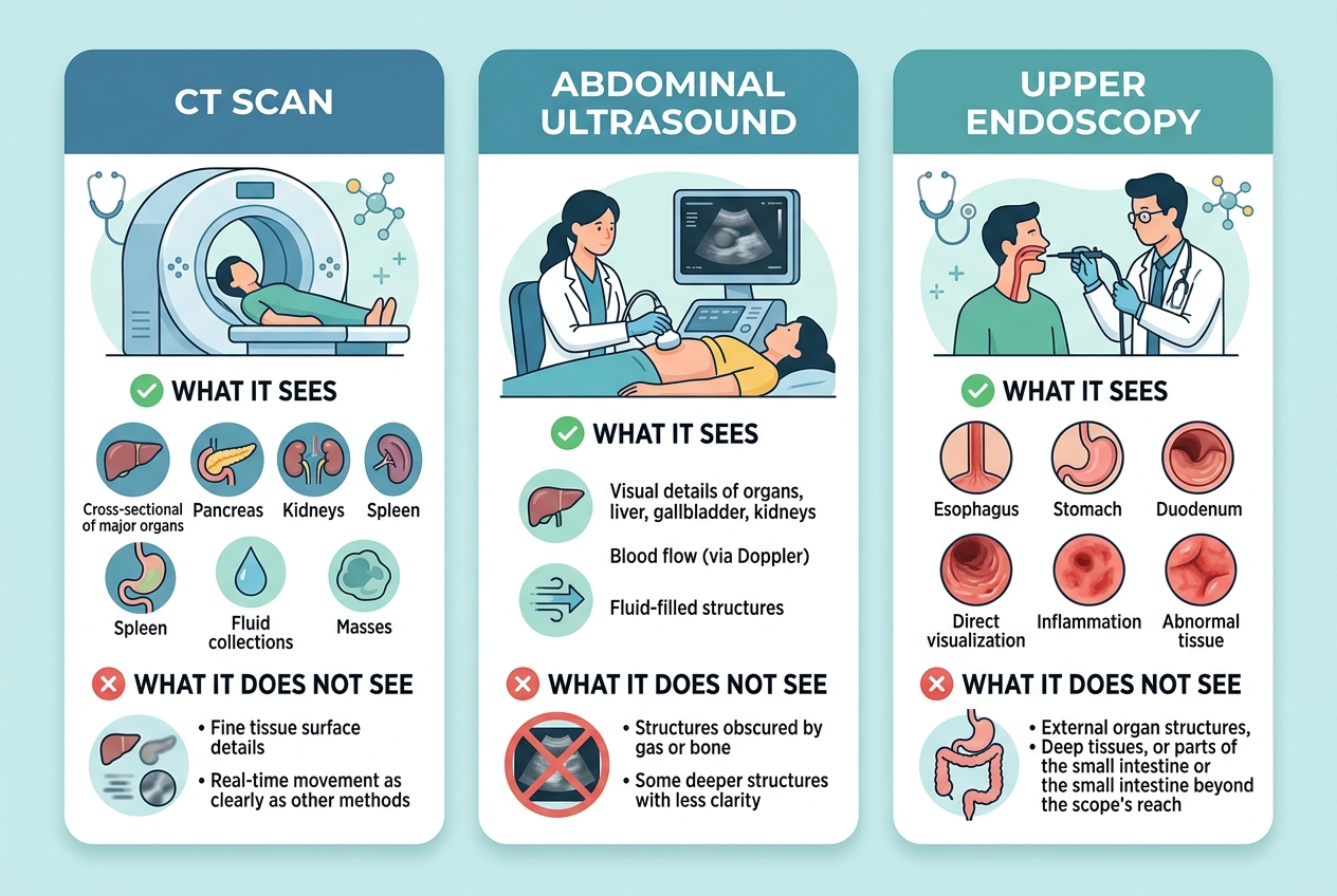
- Inline image: Clean comparison infographic with three columns labeled CT scan, abdominal ultrasound, and upper endoscopy, showing what each test sees and does not see, calm clinical colors, readable labels. Alt text: Infographic comparing CT scan, abdominal ultrasound, and upper endoscopy by what each test can evaluate.
Leave a Reply